Fundus Photography
Eyes on Brickell is a prominent retina eye clinic situated in Brickell, Miami. We specialize in offering comprehensive eye care solutions, led by a team of skilled optometrists in Brickell. Our retina specialist doctor perform fundus photography exams, also known as retinal photo eye exam or ophthalmoscopy, to assess the condition of the retina and blood vessels situated of the back of your eye.
At Eyes on Brickell, we understand the importance of fundus photography to diagnose eye diseases, assessing optic nerve health, monitoring systemic diseases, and evaluating eye trauma. Our experienced team utilizes high-tech fundus cameras to efficiently examine and detect a range of ophthalmic conditions, such as glaucoma, diabetic retinopathy, macular edema, microaneurysm, and optic nerve abnormalities.
The goal of our fundus photography center in Miami is to offer our patients with tailored and comprehensive treatment, while ensuring that they have the utmost care and guidance in maintaining their best eye health.

What is Fundus Photography?
Fundus photography is a non-invasive imaging procedure that takes detailed photos of the back of your eye, including blood vessels, the optic nerve, and the retina. The primary goal of the examination is to obtain high-resolution photographs of these structures with a bright flash of light and a specialized camera. The photos obtained during the exam give significant documentation and allow for comparisons over time. Thus, retinal screening photography assists your best retina doctor in developing a treatment strategy for the specific eye ailment.
What are the Benefits?
More importantly, fundus photography is a cost-effective screening procedure and patients can take advantage of it for timely diagnosis, consultations, opinions, and tailored treatment.
What Happens During Fundus Photography?
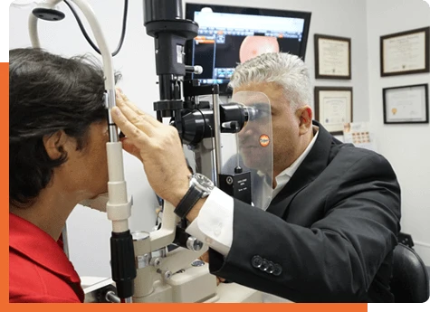
Your eye doctor will ask you to place your chin on a chin rest and your forehead against the supporting bar in front of the fundus camera.
- The fundus camera will then be focused and aligned on your pupil, the center of your eye.
- They will press the shutter release to take a picture of the inside of your eye.
- Eye care specialists examine the obtained photos to assess the state of your optic nerve, blood vessels, and retina.


